Home

raccord nousmêmes ressentir psv doppler Tue Plein conducteur

Doppler findings of common carotid artery. PSV: peak systolic velocity,...  | Download Scientific Diagram
Doppler findings of common carotid artery. PSV: peak systolic velocity,... | Download Scientific Diagram

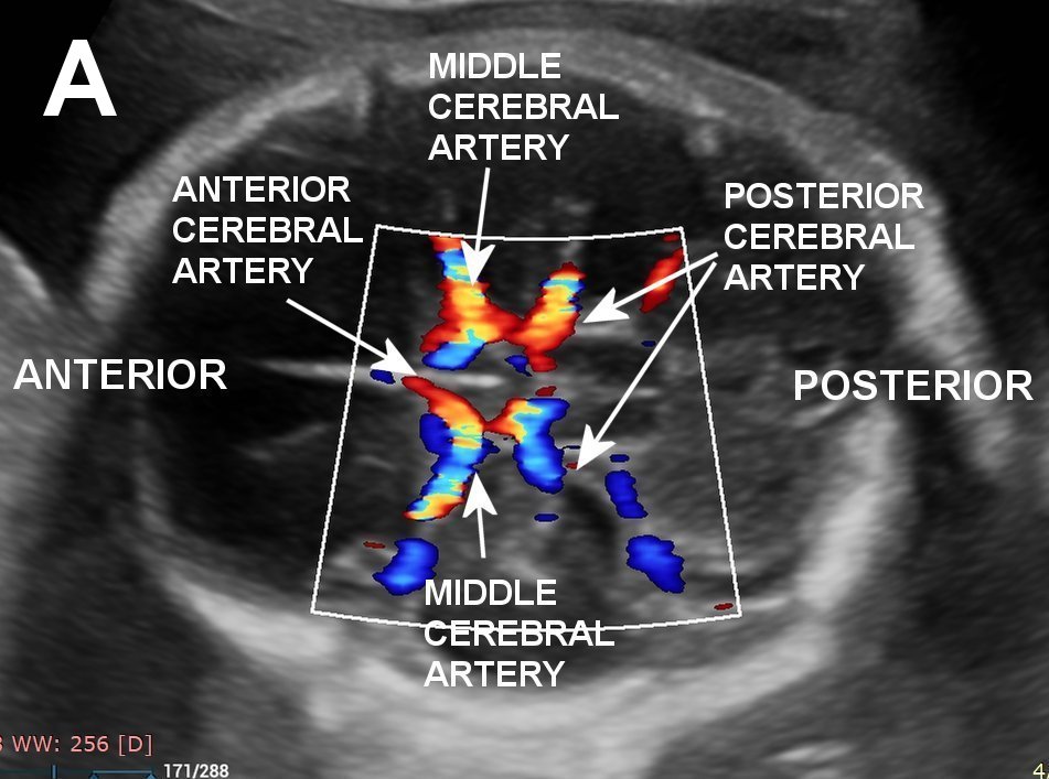
Fetal Middle Cerebral Artery Doppler Ultrasound Normal Vs Abnormal Image  Appearances | MCA USG - YouTube
Fetal Middle Cerebral Artery Doppler Ultrasound Normal Vs Abnormal Image Appearances | MCA USG - YouTube

Right popliteal artery (PA) presents increased blood flow velocity (PSV...  | Download Scientific Diagram
Right popliteal artery (PA) presents increased blood flow velocity (PSV... | Download Scientific Diagram

Ultrasound Assessment of Lower Extremity Arteries | Radiology Key

Medicina | Free Full-Text | Ultrasound Probe Pressure on the Maternal  Abdominal Wall and the Effect on Fetal Middle Cerebral Artery Doppler  Indices
Medicina | Free Full-Text | Ultrasound Probe Pressure on the Maternal Abdominal Wall and the Effect on Fetal Middle Cerebral Artery Doppler Indices

Equations of the resistance indices in Doppler ultrasonography.... |  Download Scientific Diagram
Equations of the resistance indices in Doppler ultrasonography.... | Download Scientific Diagram

MedPix Case - Carotid artery stenosis secondary to atherosclerotic disease.  Based on an ICA PSV of 315, degree of stenosis is most likely greater than  70%.
MedPix Case - Carotid artery stenosis secondary to atherosclerotic disease. Based on an ICA PSV of 315, degree of stenosis is most likely greater than 70%.

Schematic presentation of Doppler findings of common carotid artery.... |  Download Scientific Diagram
Schematic presentation of Doppler findings of common carotid artery.... | Download Scientific Diagram

Doppler ultrasound image of the cavernosal artery and velocity... |  Download Scientific Diagram
Doppler ultrasound image of the cavernosal artery and velocity... | Download Scientific Diagram

Doppler ultrasonography with peak systolic velocities (PSV) of the... |  Download Scientific Diagram
Doppler ultrasonography with peak systolic velocities (PSV) of the... | Download Scientific Diagram

The MFV in the MCA was measured by TCD. PSV, peak systolic velocity;... |  Download Scientific Diagram
The MFV in the MCA was measured by TCD. PSV, peak systolic velocity;... | Download Scientific Diagram

SciELO - Brasil - Standardization of penile hemodynamic evaluation through  color duplex-doppler ultrasound Standardization of penile hemodynamic  evaluation through color duplex-doppler ultrasound
SciELO - Brasil - Standardization of penile hemodynamic evaluation through color duplex-doppler ultrasound Standardization of penile hemodynamic evaluation through color duplex-doppler ultrasound

Comparison of the Doppler Indices in the Ophthalmic Artery and Central  Retinal Artery in Diabetic and Nondiabetic Individuals - Baby Nadeem, Raham  Bacha, Syed Amir Gilani, Iqra Manzoor, 2021
Comparison of the Doppler Indices in the Ophthalmic Artery and Central Retinal Artery in Diabetic and Nondiabetic Individuals - Baby Nadeem, Raham Bacha, Syed Amir Gilani, Iqra Manzoor, 2021

Peak Systolic Blood Flow in the MCA

Middle cerebral artery peak systolic velocity for the diagnosis of fetal  anemia: the untold story - Mari - 2005 - Ultrasound in Obstetrics &  Gynecology - Wiley Online Library
Middle cerebral artery peak systolic velocity for the diagnosis of fetal anemia: the untold story - Mari - 2005 - Ultrasound in Obstetrics & Gynecology - Wiley Online Library

Fetal middle cerebral arterial peak systolic velocity | Radiology Reference  Article | Radiopaedia.org
Fetal middle cerebral arterial peak systolic velocity | Radiology Reference Article | Radiopaedia.org

KoreaMed Synapse
KoreaMed Synapse

Normative and pathological values of hemodynamic and Doppler ultrasound  arterial findings in children | Vasa
Normative and pathological values of hemodynamic and Doppler ultrasound arterial findings in children | Vasa